Your skin barrier represents one of the most sophisticated biological defence systems in the human body, yet its complexity remains largely underappreciated by both consumers and some healthcare professionals. This remarkable structure, scientifically known as the stratum corneum, functions as a dynamic interface between your internal physiology and the external environment, orchestrating multiple protective mechanisms simultaneously. Beyond simple moisture retention, your skin barrier coordinates antimicrobial defence, pH regulation, temperature control, and selective permeability—all while maintaining structural integrity under constant environmental assault. Understanding how this intricate system operates reveals why targeted barrier support often proves more effective than symptomatic treatments for various skin conditions.
Stratum corneum architecture: understanding your skin’s primary defence mechanism
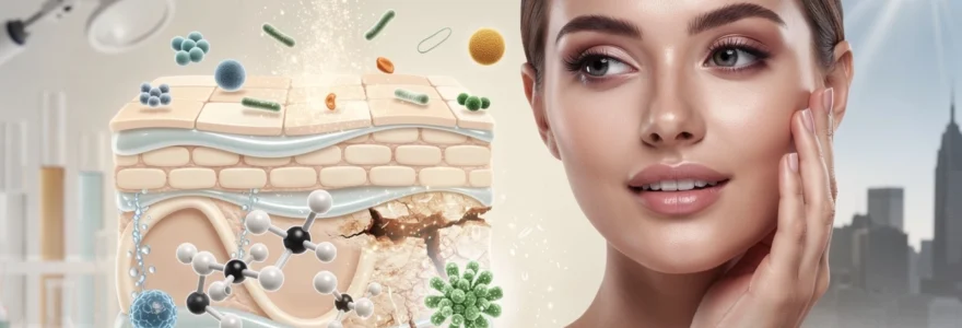
The stratum corneum represents the culmination of a precisely orchestrated cellular differentiation process that begins in the basal layer of the epidermis. This outermost skin layer consists of 15-20 rows of flattened, dead cells called corneocytes, embedded within a continuous lipid matrix. The entire structure measures approximately 10-40 micrometers in thickness—roughly equivalent to a sheet of plastic wrap—yet provides protection equivalent to a sophisticated biological fortress.
Corneocyte structure and keratinocyte differentiation process
Corneocytes originate from living keratinocytes through a remarkable transformation process called terminal differentiation. During this 14-day journey from the basal layer to the surface, keratinocytes progressively lose their organelles, flatten significantly, and develop a robust protein envelope. The mature corneocyte becomes filled primarily with keratin filaments and natural moisturising factors, creating a flexible yet resilient building block. This differentiation process responds dynamically to environmental conditions, with factors like humidity, temperature, and mechanical stress influencing corneocyte production rates and structural properties.
Lipid bilayer composition: ceramides, cholesterol, and free fatty acids
The intercellular lipid matrix surrounding corneocytes consists of three primary lipid classes arranged in highly organised lamellar structures. Ceramides comprise approximately 50% of barrier lipids, with at least 12 distinct ceramide species identified in human skin. Each ceramide type contributes specific functional properties, from moisture retention to antimicrobial activity. Cholesterol constitutes roughly 25% of barrier lipids and provides membrane fluidity regulation, whilst free fatty acids make up the remaining 25%, contributing to pH maintenance and antimicrobial defence. This precise lipid ratio proves critical for optimal barrier function, with even minor disruptions potentially compromising protective capacity.
Desmosome junction breakdown and natural desquamation cycle
The controlled breakdown of cellular connections enables natural skin renewal through desquamation—the shedding of surface corneocytes. Desmosomes, protein complexes that initially bind keratinocytes together, undergo enzymatic degradation as cells approach the skin surface. This process involves multiple proteases, including kallikreins and cathepsins, which respond to humidity levels and pH changes. Proper desquamation maintains barrier thickness whilst removing aged, potentially damaged cells. Disrupted desquamation can lead to hyperkeratosis or excessive barrier thinning, both of which compromise protective function.
Transepidermal water loss (TEWL) regulation mechanisms
TEWL regulation represents one of the barrier’s most critical functions, preventing dehydration whilst maintaining skin flexibility. Multiple mechanisms control water movement, including lipid lamellae that create tortuous pathways for water molecules, and hygroscopic natural moisturising factors that bind water within corneocytes. Healthy skin maintains TEWL rates between 4-8 g/m²/h under normal conditions. Environmental factors significantly influence TEWL, with low humidity increasing water loss and high humidity potentially reducing barrier integrity through excessive hydration. Understanding TEWL patterns helps assess barrier health and predict skin responses to environmental changes.
The skin barrier’s ability to maintain optimal hydration levels whilst protecting against external threats represents millions of years of evolutionary refinement, creating a system more sophisticated than any current technological equivalent.
Filaggrin protein function and barrier integrity maintenance
Natural moisturising factor (NMF) production and amino acid conversion
Filaggrin begins its life as a large precursor protein called profilaggrin, stored within keratohyalin granules in the granular layer of the epidermis. As keratinocytes move toward the skin surface and undergo terminal differentiation, profilaggrin is enzymatically cleaved into multiple filaggrin units. These filaggrin molecules bind and aggregate keratin filaments, causing the cells to flatten and form the dense, mechanically resilient corneocytes that characterise a healthy skin barrier.
Once filaggrin has completed its structural role, it is further broken down into a range of free amino acids and their derivatives. Collectively known as natural moisturising factor, or NMF, these hygroscopic compounds—including pyrrolidone carboxylic acid (PCA), urocanic acid, lactic acid, and various salts—act like microscopic sponges within the corneocyte. By binding and holding water, NMF maintains optimal corneocyte hydration, elasticity, and enzyme function, directly influencing how smooth and comfortable your skin feels on a daily basis.
Filaggrin gene mutations: FLG variants and atopic dermatitis correlation
Filaggrin production is controlled by the FLG gene, and loss-of-function mutations in this gene represent one of the most robustly documented risk factors for atopic dermatitis (eczema). Individuals carrying certain FLG variants produce less filaggrin or truncated forms of the protein, leading to reduced NMF levels and impaired barrier cohesion. The result is increased transepidermal water loss, microfissuring of the stratum corneum, and easier penetration of allergens and irritants.
Multiple population studies suggest that up to 30–50% of people with moderate to severe atopic dermatitis carry at least one filaggrin mutation. Whilst not everyone with an FLG variant develops eczema, having this genetic predisposition significantly lowers the threshold for barrier disruption when combined with environmental stressors. For affected individuals, this means that maintaining barrier integrity through gentle cleansing, regular emollient use, and early intervention during flares is not just helpful—it is central to long-term disease control.
Ph regulation through filaggrin-derived histidine metabolism
The skin surface maintains a slightly acidic pH—typically between 4.5 and 5.5—often referred to as the acid mantle. This acidity is crucial for optimal enzyme activity in the stratum corneum, healthy microbial balance, and efficient barrier repair. Filaggrin degradation contributes directly to this pH regulation. One of its component amino acids, histidine, is converted into urocanic acid, which helps acidify the stratum corneum and stabilise the skin barrier’s chemical environment.
When filaggrin levels are reduced—whether through genetic variants, chronic inflammation, or excessive cleansing—the downstream production of histidine-derived acids diminishes. This can shift the skin surface toward a more alkaline state, impairing desquamation enzymes, altering lipid-processing pathways, and encouraging colonisation by less desirable microbes. In practice, this is one reason why harsh, high-pH cleansers can be so disruptive: they undermine both the physical components of the barrier and its finely tuned pH balance.
Seasonal humidity impact on filaggrin expression levels
Filaggrin expression is not fixed; it responds to environmental conditions, particularly ambient humidity. In low-humidity climates or during cold, dry seasons, filaggrin production and subsequent NMF levels can decrease, exacerbating dryness and sensitivity. This is one reason many people experience itching, flaking, or eczema flares in winter—even if their routine has not changed. The barrier is simply under greater stress and has fewer hydrating resources available.
Conversely, higher humidity environments can support more robust filaggrin processing and slightly improved NMF content, often translating into softer, more comfortable skin. However, this does not mean humidity alone can compensate for aggressive skincare habits. From a practical standpoint, adjusting your routine seasonally—using richer moisturisers and more occlusive textures in dry months, and maintaining consistent use of barrier-supportive ingredients year-round—helps buffer these humidity-driven shifts in filaggrin activity.
Microbial ecosystem balance and antimicrobial peptide production
Your skin barrier is not a sterile shield; it is a living ecosystem populated by billions of microorganisms. This community—bacteria, yeasts, and viruses collectively known as the skin microbiome—interacts constantly with the physical barrier to maintain health. Far from being passive passengers, many of these microbes produce metabolites and antimicrobial compounds that reinforce your barrier’s defence capabilities. When this microbial balance is disturbed, or dysbiosis develops, the risk of inflammation, infection, and chronic skin conditions rises significantly.
The skin itself contributes to microbial regulation through the production of antimicrobial peptides (AMPs), such as defensins and cathelicidins. These short proteins act like targeted antibiotics produced on demand, selectively inhibiting potential pathogens whilst generally sparing beneficial residents. Barrier integrity, pH, and lipid composition all influence which microbes thrive, meaning that supporting your skin barrier indirectly supports a balanced microbiome—and vice versa.
Staphylococcus epidermidis colonisation and beneficial metabolite synthesis
One of the most important commensal bacteria on human skin is Staphylococcus epidermidis. Under healthy conditions, this organism colonises the stratum corneum and hair follicles, forming a kind of microbial “neighbourhood watch.” S. epidermidis produces a range of metabolites, including short-chain fatty acids and antimicrobial peptides, that inhibit the growth of more aggressive species such as Staphylococcus aureus. Some strains even stimulate host AMP production, enhancing the skin’s innate immunity.
When your barrier is intact, sebum composition appropriate, and pH balanced, S. epidermidis tends to flourish in a way that benefits you. However, harsh antiseptics, overuse of antibacterial cleansers, or prolonged antibiotic therapy can reduce its numbers, opening ecological space for less desirable microbes. This is one reason why “over-sanitising” the skin can paradoxically lead to more infections or irritation; you are weakening the very ecosystem that helps defend you.
Defensin and cathelicidin antimicrobial peptide mechanisms
Antimicrobial peptides represent one of the skin barrier’s most elegant defence tools. Defensins and cathelicidins (such as LL-37) are small, positively charged molecules that insert into microbial membranes, creating pores that ultimately lead to cell death. Unlike traditional antibiotics, AMPs act rapidly and broadly, targeting bacteria, some fungi, and certain viruses. Their production is upregulated in response to injury, infection, and specific microbial signals.
AMP activity is closely tied to barrier status. Adequate vitamin D, balanced pH, and intact lipid structures all support appropriate peptide expression and function. When the barrier is chronically inflamed or disrupted, AMP production can become dysregulated—either insufficient, allowing opportunistic infections, or excessive, contributing to inflammatory skin diseases. Supporting barrier health therefore helps stabilise this antimicrobial “security system,” ensuring it reacts proportionately rather than erratically.
Malassezia furfur overgrowth and seborrhoeic dermatitis development
Not all microorganisms are strictly beneficial or harmful; many, like Malassezia furfur, occupy a grey zone where context determines their role. Malassezia species are lipid-dependent yeasts that form part of the normal skin flora, especially in sebum-rich areas such as the scalp, face, and chest. Under balanced conditions, they coexist quietly with the barrier. However, when barrier lipids shift in composition, humidity is high, or immune responses become dysregulated, these yeasts can overgrow.
This overgrowth is strongly associated with seborrhoeic dermatitis and dandruff, conditions characterised by flaky, sometimes itchy, inflamed skin. The yeasts metabolise sebum triglycerides into free fatty acids that can irritate the stratum corneum, further compromising barrier function. From a management perspective, combining gentle barrier-repair strategies with targeted antifungal agents often proves more effective than focusing solely on symptom relief, because you are addressing both the microbial imbalance and the underlying barrier vulnerability.
Propionibacterium acnes dysbiosis in compromised barrier states
Cutibacterium acnes (historically known as Propionibacterium acnes) is another resident microbe whose behaviour shifts with barrier status. In healthy skin, it helps maintain an acidic environment and metabolises sebum components, contributing to normal physiology. In compromised barrier states, especially when sebum production is high and follicular openings become obstructed, certain strains of C. acnes proliferate excessively and trigger inflammatory cascades associated with acne.
Emerging research suggests that acne is not simply a matter of “too much bacteria,” but rather an imbalance between different C. acnes subtypes and a barrier environment that favours inflammation. Harsh, drying treatments that strip lipids and disrupt the acid mantle may temporarily reduce oiliness, but they can also worsen dysbiosis and prolong recovery. A more sustainable strategy pairs evidence-based acne therapies with barrier-supportive practices—non-comedogenic moisturisers, pH-appropriate cleansers, and gradual introduction of actives—to help rebalance the follicular microbiome.
Environmental stressor impact on barrier function recovery
Every day, your skin barrier navigates a barrage of environmental stressors that can slow or even reverse its natural repair processes. Ultraviolet radiation generates reactive oxygen species that damage barrier lipids and proteins, accelerating transepidermal water loss and undermining structural cohesion. Air pollution adds another layer of oxidative and inflammatory stress, as fine particles and chemical pollutants adhere to the stratum corneum, disrupt lipids, and may penetrate through impaired areas of the barrier.
Climate factors—temperature extremes, low humidity, high wind, and air conditioning—further influence how quickly the barrier can recover after insult. For instance, controlled studies show that exposure to low humidity environments can increase TEWL by more than 50%, prolonging recovery times after even mild irritation. What does this mean in practical terms? If you are trying to repair a damaged barrier but spending long hours in dry, heated or air-conditioned spaces without compensatory skincare, your progress may plateau or feel frustratingly slow.
Modifiable lifestyle factors also play a role. Sleep deprivation has been linked to delayed barrier recovery after tape stripping in experimental settings, likely due to altered hormone profiles and reduced nocturnal repair activity. Psychological stress can increase cortisol levels, which, in turn, impair lipid synthesis and amplify inflammation. To support barrier function recovery, topical measures—like ceramide-rich moisturisers and daily sunscreen—work best when paired with environmental adjustments: using humidifiers in dry environments, avoiding unnecessary sun exposure, and building habits that support adequate rest.
Clinical assessment methods for barrier dysfunction diagnosis
Accurately assessing barrier integrity allows clinicians to distinguish between primary barrier disorders and secondary barrier involvement in broader dermatological conditions. One widely used quantitative method is transepidermal water loss measurement with an open-chamber or closed-chamber evaporimeter. By capturing the water vapour gradient over the skin, these instruments provide objective TEWL values that help identify subclinical barrier impairment and track response to treatment over time.
Another informative tool is corneometry, which evaluates stratum corneum hydration via changes in skin capacitance. Low corneometer readings often correlate with reduced NMF levels and disrupted lipid structures. In combination with TEWL data, corneometry helps build a nuanced picture of whether the barrier is primarily leaky, under-hydrated, or both. More advanced techniques—such as confocal microscopy or tape stripping followed by biochemical analysis—can assess corneocyte cohesion, lipid organisation, and inflammatory markers, but these are typically reserved for research or specialised clinics.
In routine practice, careful visual and tactile examination remains indispensable. Dermatologists assess scale, erythema, glossiness, and surface texture, alongside patient-reported symptoms like stinging, burning, or disproportionate reactions to skincare products. Simple clinical tests, such as observing how skin responds to a mild surfactant patch or how quickly erythema resolves after minor irritation, can offer additional clues about barrier resilience. When needed, patch testing helps differentiate allergic contact dermatitis from barrier-driven sensitivity, ensuring that treatment focuses on the correct underlying mechanism.
Targeted barrier repair strategies using ceramide-dominant formulations
Ceramide-dominant formulations are specifically designed to replenish the lipid “mortar” that holds your corneocyte “bricks” together, making them a cornerstone of modern barrier repair strategies. By combining ceramides with cholesterol and free fatty acids in ratios that approximate those found in healthy skin, these formulations support the re-establishment of organised lamellar structures. Clinical studies have shown that consistent use of such products can reduce TEWL, increase hydration, and improve visible signs of dryness and irritation within a few weeks.
To maximise benefits, ceramide-focused moisturisers should be applied to slightly damp skin after gentle cleansing, ideally within a few minutes of stepping out of the shower or washing your face. This timing allows the product to trap existing surface moisture while delivering lipids into the upper layers of the stratum corneum. In more severely compromised barriers—such as in atopic dermatitis or after procedural treatments—layering a ceramide cream under a more occlusive ointment at night can create a “moisture sandwich” that accelerates recovery.
Of course, even the most sophisticated ceramide-dominant product cannot fully compensate for ongoing barrier insults. For meaningful, sustained improvement, you need to pair targeted formulations with a barrier-conscious routine: avoiding harsh surfactants and over-exfoliation, selecting pH-appropriate products, and maintaining year-round UV protection. When you combine these behavioural shifts with evidence-based barrier repair ingredients, you give your stratum corneum the best possible conditions to do what it has evolved to do—protect you, quietly and efficiently, every hour of the day.
 Good health cannot be bought, but rather is an asset that you must create and then maintain on a daily basis.
