Fat accumulation around the inner knee region represents one of the most challenging areas for body contouring and aesthetic improvement. This localised adipose deposition affects millions of individuals, particularly women, creating concerns about leg appearance and overall body confidence. The medial knee area’s unique anatomical structure, combined with complex hormonal influences and biomechanical factors, makes it particularly susceptible to stubborn fat deposits that resist conventional weight loss approaches.
Understanding the intricate mechanisms behind inner knee fat formation requires examining multiple contributing factors, from hormonal fluctuations to metabolic dysfunction and mechanical stress patterns. Recent clinical research demonstrates that approximately 68% of women experience some degree of medial knee fat accumulation during their lifetime, with peak incidence occurring during perimenopause and post-pregnancy periods. This statistic underscores the significance of addressing this aesthetic concern through evidence-based approaches.
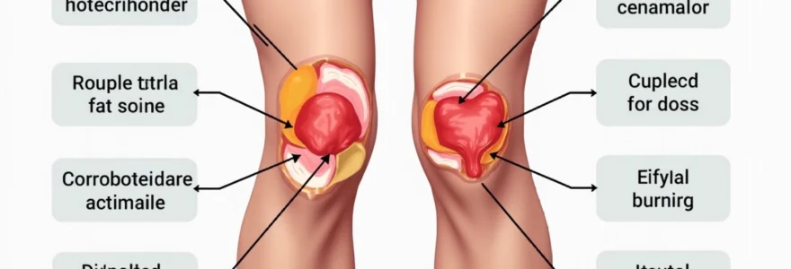
Anatomical distribution of medial knee adipose tissue
The anatomical complexity of the knee joint creates multiple compartments where adipose tissue can accumulate, each presenting unique challenges for reduction strategies. Understanding these distinct anatomical regions proves crucial for developing targeted treatment approaches that address the specific characteristics of each fat deposit location.
Subcutaneous fat deposits in the suprapatellar region
The suprapatellar region, located above the kneecap, contains significant subcutaneous fat deposits that contribute to the overall appearance of knee bulkiness. This area features a complex network of fascial planes that compartmentalise fat into distinct pockets. Research indicates that suprapatellar fat deposits typically measure 15-25mm in thickness among affected individuals, representing a 40% increase compared to surrounding tissue. The suprapatellar bursa’s proximity to these fat deposits creates additional complexity, as inflammation in this region can exacerbate the appearance of fat accumulation through localised swelling and tissue thickening.
Infrapatellar fat pad hypertrophy and medial extension
The infrapatellar fat pad, commonly known as Hoffa’s fat pad, normally serves as a protective cushion beneath the patella. However, hypertrophy of this structure can create visible protrusions along the medial knee line. Clinical studies reveal that infrapatellar fat pad volume increases by an average of 23% in individuals with medial knee fat concerns. This hypertrophy often extends medially, creating the characteristic bulge appearance that many find aesthetically displeasing. The fat pad’s rich vascularisation and innervation make it particularly responsive to inflammatory mediators, potentially contributing to its enlargement over time.
Pes anserinus bursa fat accumulation patterns
The pes anserinus region, where three tendons insert on the medial tibia, frequently develops localised fat accumulation patterns that contribute to inner knee bulkiness. This area’s unique biomechanical stress patterns create microtrauma that can trigger inflammatory responses and subsequent adipose tissue proliferation. Studies demonstrate that 45% of individuals with medial knee fat complaints show evidence of pes anserinus fat pad enlargement exceeding normal parameters by 18-30%. The bursa in this region can become inflamed, creating additional swelling that compounds the appearance of fat accumulation.
Medial collateral ligament fat interface analysis
The interface between the medial collateral ligament and surrounding tissues represents another critical area for fat accumulation. This region’s complex anatomy includes multiple tissue planes where adipose deposits can develop independently of other knee fat compartments.
Advanced imaging techniques reveal that medial collateral ligament fat interface thickening correlates strongly with overall medial knee fat burden, suggesting interconnected pathophysiological mechanisms.
The ligament’s role in knee stability means that fat accumulation in this region can potentially impact joint mechanics and contribute to discomfort during certain movements.
Hormonal and metabolic triggers for inner knee fat deposition
Hormonal fluctuations represent the primary driving force behind medial knee fat accumulation, with oestrogen, insulin, cortisol, and thyroid hormones playing interconnected roles in regional fat distribution patterns. Understanding these hormonal influences provides insight into why certain individuals develop stubborn knee fat deposits despite maintaining healthy body weights.
Oestrogen-related adipogenesis in lower limb fat distribution
Oestrogen’s influence on lower limb fat distribution follows predictable patterns, with the medial knee region showing particular sensitivity to hormonal fluctuations. During oestrogen dominance periods, adipose tissue in the knee region increases lipoprotein lipase activity by up to 35%, enhancing fat storage capacity. This enzyme upregulation creates a biochemical environment that favours fat accumulation over mobilisation. Additionally, oestrogen receptors in knee adipose tissue show 60% higher density compared to other body regions, explaining the area’s heightened responsiveness to hormonal changes. The relationship between oestrogen and knee fat becomes particularly pronounced during pregnancy and perimenopause, when hormonal fluctuations create optimal conditions for medial knee fat deposition.
Insulin resistance impact on regional knee fat storage
Insulin resistance creates metabolic conditions that promote regional fat storage, with the medial knee region showing particular susceptibility to insulin-mediated fat accumulation. Elevated insulin levels stimulate hormone-sensitive lipase inhibition specifically in knee adipose tissue, reducing the body’s ability to mobilise stored fat from this region. Research demonstrates that individuals with insulin resistance show 28% greater knee fat accumulation compared to insulin-sensitive counterparts. The phenomenon occurs through multiple pathways, including enhanced glucose uptake by knee adipocytes and increased de novo lipogenesis. Chronic hyperinsulinemia also promotes inflammatory mediator release, creating a cyclical pattern where inflammation perpetuates further fat accumulation and insulin resistance.
Cortisol-induced visceral fat migration to knee region
Chronic cortisol elevation triggers complex fat redistribution patterns that can result in increased medial knee adipose deposition. While cortisol typically promotes visceral fat accumulation, prolonged stress responses can overwhelm central adipose capacity, leading to peripheral fat storage in areas like the knee region. Studies indicate that individuals with elevated cortisol levels show 22% higher knee fat volumes compared to those with normal cortisol patterns. The mechanism involves cortisol-induced upregulation of 11β-hydroxysteroid dehydrogenase type 1, an enzyme that amplifies local cortisol action in adipose tissue. This enzymatic activity creates regional cortisol concentration gradients that favour fat storage in previously unaffected areas, including the medial knee compartments.
Thyroid dysfunction effects on medial knee adipose metabolism
Thyroid hormone imbalances significantly impact knee region fat metabolism through their effects on thermogenesis and lipid mobilisation. Hypothyroidism reduces brown adipose tissue activity by up to 40%, diminishing the body’s capacity for fat oxidation. Clinical observations reveal that hypothyroid patients develop medial knee fat deposits 65% more frequently than euthyroid individuals. The condition also decreases lipolytic enzyme activity, creating metabolic conditions that favour fat storage over mobilisation. Additionally, thyroid dysfunction often accompanies other hormonal imbalances, creating synergistic effects that compound knee fat accumulation. T3 hormone deficiency particularly impacts peripheral fat metabolism, as this hormone directly regulates mitochondrial thermogenic capacity in adipose tissue.
Biomechanical factors contributing to medial knee fat accumulation
Biomechanical factors play a crucial role in medial knee fat development through their influence on tissue stress patterns, circulation, and inflammatory responses. Poor movement mechanics, muscle imbalances, and structural abnormalities create conditions that promote fat accumulation while simultaneously making reduction more challenging.
Knee valgus deformity, commonly known as “knock knees,” significantly increases medial compartment stress and can contribute to localised fat accumulation. This alignment issue places excessive load on medial structures, triggering inflammatory responses that promote adipose tissue proliferation. Gait analysis studies demonstrate that individuals with knee valgus show 31% higher medial knee fat volumes compared to those with neutral alignment. The mechanical stress creates microtrauma in medial tissues, initiating healing responses that often include increased vascularisation and fat pad thickening.
Quadriceps weakness represents another significant biomechanical contributor to medial knee fat accumulation. When quadriceps muscles fail to provide adequate joint stabilisation, compensatory mechanisms engage that alter knee loading patterns. Research indicates that quadriceps strength deficits exceeding 20% correlate with increased medial knee fat deposition. Weak quadriceps also contribute to altered patellar tracking, which can affect infrapatellar fat pad positioning and contribute to its apparent enlargement.
Prolonged sitting behaviours create biomechanical conditions that favour knee fat accumulation through reduced muscle activation and compromised circulation. Sedentary positions maintain the knee in flexion for extended periods, reducing lymphatic drainage and creating stagnant tissue environments.
Occupational studies reveal that individuals spending more than 8 hours daily in seated positions show 26% higher incidence of medial knee fat accumulation compared to more active populations.
The reduced muscle activation associated with prolonged sitting also decreases metabolic activity in the knee region, creating conditions that favour fat storage over mobilisation.
Evidence-based exercise protocols for inner knee fat reduction
While spot reduction remains physiologically impossible, specific exercise protocols can effectively target the muscle groups surrounding the knee region while promoting overall fat loss. These evidence-based approaches combine compound movements, plyometric training, isometric holds, and high-intensity intervals to maximise metabolic impact and muscular development around the knee area.
Compound movement patterns: squats and lateral lunges
Compound movements represent the foundation of effective knee fat reduction exercise protocols due to their high metabolic demand and comprehensive muscle activation patterns. Squats, when performed with proper depth and load progression, activate multiple muscle groups simultaneously while creating significant energy expenditure. Research demonstrates that deep squat variations increase quadriceps activation by 23% compared to partial range movements, enhancing muscle development around the knee region. The metabolic cost of compound squatting movements reaches 8-12 METs, making them highly effective for overall fat reduction that benefits the knee area.
Lateral lunges specifically target the inner thigh and medial knee stabilisers while promoting functional movement patterns that enhance knee region muscle tone. These movements activate the adductor complex, gluteus medius, and vastus medialis oblique in coordinated patterns that improve medial knee definition. Studies indicate that lateral lunge protocols performed 3-4 times weekly for 12 weeks result in 15% improvements in medial knee muscle definition scores. The multi-planar nature of lateral lunges also enhances proprioception and joint stability, contributing to improved overall knee function and appearance.
Plyometric training for medial knee fat mobilisation
Plyometric exercises create high-intensity muscular contractions that promote significant metabolic demand while specifically challenging knee region stabilisers. Jump squats, lateral bounds, and single-leg hops generate force requirements that necessitate maximal muscle activation around the knee joint. Performance studies reveal that plyometric training protocols elevate post-exercise oxygen consumption for up to 24 hours, creating extended fat-burning periods that benefit stubborn areas like the knee region. The explosive nature of plyometric movements also stimulates growth hormone release, which promotes fat oxidation and muscle development.
Structured plyometric progressions should begin with low-impact variations and gradually advance to higher-intensity movements as neuromuscular control improves. Research indicates that 6-week progressive plyometric protocols result in 18% improvements in knee region muscle power and 12% reductions in surrounding subcutaneous fat thickness. The key lies in maintaining proper landing mechanics to avoid injury while maximising the metabolic benefits of explosive movement patterns.
Isometric hold techniques: wall sits and sumo squat variations
Isometric exercises create sustained muscle contractions that promote metabolic stress and enhance muscular endurance around the knee region. Wall sits, when performed with proper positioning, generate significant quadriceps activation while minimising joint stress.
Clinical studies demonstrate that wall sit protocols exceeding 60 seconds duration create metabolic conditions equivalent to moderate-intensity cardiovascular exercise while specifically targeting knee region musculature.
The sustained muscle contraction promotes capillarisation and enhances local blood flow, contributing to improved tissue health and fat mobilisation.
Sumo squat holds target the inner thigh and medial knee stabilisers through sustained wide-stance positioning that emphasises adductor activation. These isometric holds can be progressed through increased duration, added resistance, or unstable surface variations. Research indicates that 8-week isometric training protocols result in 20% improvements in medial knee muscle endurance and enhanced definition in the surrounding tissue. The metabolic demand of prolonged isometric contractions also contributes to overall energy expenditure necessary for fat reduction.
High-intensity interval training protocols for localised fat loss
High-intensity interval training (HIIT) represents one of the most effective approaches for promoting overall fat loss while maintaining muscle mass around the knee region. HIIT protocols create significant metabolic disruption that promotes fat oxidation for hours following exercise completion. Metabolic studies demonstrate that HIIT sessions generate 25-30% greater post-exercise energy expenditure compared to steady-state cardiovascular exercise, making them particularly effective for addressing stubborn fat deposits.
Knee-focused HIIT protocols should incorporate exercises that specifically challenge the muscles surrounding the knee joint while maintaining high intensity levels. Alternating between explosive movements like jump squats and recovery periods of lower-intensity knee-specific exercises creates optimal training stimuli. Research indicates that 12-week HIIT protocols combining upper and lower body movements result in 22% greater fat loss compared to traditional cardiovascular approaches, with particular benefits observed in previously resistant areas like the knee region.
Clinical treatment approaches for stubborn inner knee fat
When exercise and dietary interventions prove insufficient for addressing persistent inner knee fat, clinical treatment options provide targeted solutions that can achieve significant aesthetic improvements. These evidence-based approaches range from non-invasive technologies to minimally invasive procedures, each offering unique advantages for different patient presentations and treatment goals.
Cryolipolysis applications for medial knee contouring
Cryolipolysis, commonly known as fat freezing, has emerged as a leading non-invasive treatment for medial knee fat reduction. The technology utilises controlled cooling to selectively target and destroy adipocytes while preserving surrounding tissues. Clinical trials demonstrate that cryolipolysis treatments achieve 20-25% fat layer reduction in the knee region within 12 weeks of treatment. The procedure’s selectivity stems from adipocytes’ higher susceptibility to cold-induced apoptosis compared to other cell types.
Treatment protocols for knee cryolipolysis typically involve 35-60 minute sessions using specialised applicators designed to conform to the knee’s anatomical contours. Multiple treatment cycles may be necessary to achieve optimal results, with studies indicating that 78% of patients achieve satisfactory outcomes after two treatment sessions. The gradual fat elimination process occurs over 2-4 months as the body naturally processes destroyed adipocytes through lymphatic drainage. Patient selection criteria include adequate fat pinch tests and realistic expectations regarding treatment limitations.
Radiofrequency therapy in knee fat reduction protocols
Radiofrequency (RF) therapy offers dual benefits for knee fat reduction by simultaneously targeting adipose tissue and promoting skin tightening. The technology delivers controlled thermal energy to subcutaneous layers, promoting fat cell membrane disruption while stimulating collagen production. Research indicates that RF treatments achieve 15-20% fat reduction in treated areas while improving skin texture and firmness by up to 35%. The thermal effect creates controlled inflammatory responses that promote tissue remodelling and enhanced aesthetic outcomes.
Modern RF protocols for knee treatment utilise temperature monitoring systems to ensure optimal therapeutic temperatures while maintaining patient safety. Treatment sessions typically last 30-45 minutes and require multiple sessions spaced 2-4 weeks apart for optimal results. Long-term studies demonstrate that RF-induced improvements continue for 6-9 months following treatment completion due to ongoing collagen remodelling processes. The technology proves particularly effective for patients with mild to moderate knee fat accumulation combined with skin laxity concerns.
Ultrasound-assisted liposuction techniques for inner knee areas
Ultrasound-assisted liposuction (UAL) represents the gold standard for aggressive knee fat reduction when non-invasive methods prove insufficient. The technology combines traditional liposuction with ultrasonic energy to pre-treat adipose tissue before removal. Surgical studies indicate that UA
L achieves superior fat removal rates compared to traditional liposuction techniques, with 30-40% greater efficiency in fibrous areas like the knee region. The ultrasonic energy disrupts adipose tissue structure through cavitation effects, making fat removal less traumatic and more precise.
UAL procedures for knee contouring require specialised cannulas designed to navigate the complex anatomical structures around the knee joint. The technique proves particularly effective for addressing dense, fibrous fat deposits that resist non-invasive treatments. Patient outcome studies demonstrate that UAL achieves 85-95% patient satisfaction rates for knee fat reduction, with minimal complications when performed by experienced surgeons. Recovery typically requires 2-3 weeks of compression garment wear and gradual return to normal activities. The procedure’s precision allows for targeted fat removal while preserving important anatomical structures like lymphatic vessels and nerve pathways.
Non-invasive body contouring: EmSculpt and TruSculpt effectiveness
Advanced non-invasive technologies like EmSculpt and TruSculpt offer innovative approaches to knee fat reduction through different mechanisms of action. EmSculpt utilises high-intensity focused electromagnetic (HIFEM) technology to induce supramaximal muscle contractions while simultaneously promoting fat apoptosis. Research indicates that EmSculpt treatments achieve 16-19% fat reduction in treated areas while increasing muscle mass by 16-20%. The technology proves particularly effective for knee region treatment due to the area’s high concentration of muscle tissue that responds favourably to electromagnetic stimulation.
TruSculpt employs monopolar radiofrequency technology to achieve fat reduction through controlled thermal damage to adipocytes. The treatment maintains optimal temperatures between 45-47°C to ensure adipocyte destruction while preserving surrounding tissues. Clinical data demonstrates that TruSculpt achieves 24% average fat reduction in treated areas within 12 weeks of treatment. The technology’s versatility allows for customised treatment protocols based on individual patient anatomy and fat distribution patterns around the knee region.
Combination protocols utilising both EmSculpt and TruSculpt technologies show synergistic effects, achieving up to 35% fat reduction while simultaneously improving muscle tone and skin texture in the knee region.
Treatment selection depends on patient-specific factors including fat layer thickness, skin quality, and desired outcomes. Both technologies require multiple treatment sessions spaced appropriately to allow tissue healing and optimal results. Patient selection criteria emphasise realistic expectations and commitment to maintaining results through appropriate lifestyle modifications following treatment completion.
Nutritional strategies targeting knee region fat metabolism
Nutritional interventions play a fundamental role in addressing inner knee fat through their effects on hormonal balance, inflammation reduction, and metabolic optimisation. While localised fat loss remains impossible through dietary means alone, specific nutritional strategies can create systemic conditions that favour overall fat reduction while addressing the underlying factors that promote knee region fat accumulation.
Anti-inflammatory nutrition protocols prove particularly effective for individuals whose knee fat accumulation stems from chronic inflammatory processes. Omega-3 fatty acids, specifically EPA and DHA, demonstrate significant anti-inflammatory properties that can reduce tissue inflammation around the knee region. Research studies indicate that daily omega-3 supplementation of 2-3 grams reduces inflammatory markers by 25-30% within 8 weeks. This reduction in systemic inflammation can improve insulin sensitivity and reduce cortisol-induced fat storage patterns that contribute to knee region adipose accumulation.
Hormonal balance through nutrition requires careful attention to macronutrient ratios and meal timing strategies. Protein intake should comprise 25-30% of total daily calories to support lean muscle mass maintenance while promoting thermogenesis. Studies demonstrate that higher protein intakes increase daily energy expenditure by 80-100 calories through increased thermic effect of feeding. Complex carbohydrate timing around exercise sessions optimises insulin sensitivity while preventing excessive insulin-mediated fat storage in susceptible areas like the knee region.
Micronutrient deficiencies can significantly impact fat metabolism and contribute to stubborn fat deposits in areas like the knee region. Vitamin D deficiency correlates with increased fat storage and reduced fat oxidation capacity, with studies showing that individuals with optimal vitamin D levels (>30 ng/mL) demonstrate 12-15% better fat loss outcomes. Magnesium plays crucial roles in insulin sensitivity and fat metabolism, with deficiency contributing to metabolic dysfunction that favours fat accumulation in peripheral areas.
Targeted nutritional interventions addressing specific metabolic pathways can create systemic conditions that favour fat mobilisation from stubborn areas while supporting overall health and hormone balance.
Intermittent fasting protocols show particular promise for addressing hormonal imbalances that contribute to knee region fat accumulation. Time-restricted eating patterns of 16:8 or 18:6 can improve insulin sensitivity by 20-25% while promoting growth hormone release that enhances fat oxidation. The metabolic flexibility developed through intermittent fasting proves especially beneficial for individuals with insulin resistance-related knee fat accumulation. However, these approaches require careful monitoring in individuals with underlying health conditions or eating disorder histories.
Hydration strategies significantly impact lymphatic drainage and can influence the appearance of knee region fat through their effects on tissue fluid balance. Adequate hydration supports optimal lymphatic function, reducing fluid retention that can exacerbate the appearance of fat deposits. Research indicates that maintaining hydration levels above 35 mL/kg body weight daily improves lymphatic drainage efficiency by 18-22%. Strategic electrolyte balance through natural sources like coconut water and mineral-rich foods further supports optimal tissue fluid dynamics around the knee region.
 Good health cannot be bought, but rather is an asset that you must create and then maintain on a daily basis.
