Foot drop affects millions of people worldwide, transforming simple daily activities like walking up stairs or crossing the street into significant challenges. This neurological condition, characterised by difficulty lifting the front part of the foot, stems from various causes including stroke, spinal injuries, peripheral nerve damage, and neurological conditions. Yet behind the clinical definitions and medical terminology lie powerful stories of determination, adaptation, and remarkable recovery.
These real-world experiences demonstrate that whilst foot drop presents substantial obstacles, innovative treatments and dedicated rehabilitation can lead to meaningful functional improvements. From breakthrough surgical interventions to cutting-edge rehabilitation technologies, patients are discovering new pathways to mobility and independence that seemed impossible just years ago.
Neurological foot drop recovery: Post-Stroke patient transformations
Stroke-related foot drop represents one of the most common causes of this condition, affecting approximately 20% of stroke survivors. The journey back to mobility often requires months of intensive rehabilitation, but emerging evidence shows that targeted interventions can produce remarkable outcomes even years after the initial injury.
Recent studies indicate that functional electrical stimulation therapy has shown success rates of up to 80% in improving dorsiflexion strength among stroke patients with foot drop. This technology works by delivering precisely timed electrical impulses to weakened muscles, essentially retraining the neural pathways that control foot movement.
Sarah mitchell’s L5 radiculopathy recovery through functional electrical stimulation
Sarah’s experience with L5 radiculopathy-induced foot drop began following a herniated disc that compressed her nerve root. Initially unable to lift her left foot, she struggled with the constant fear of tripping and the exhausting cognitive load that walking demanded.
The mental effort required just to walk safely was overwhelming. Every step required conscious thought about foot placement and balance.
After six months of traditional physiotherapy showed limited progress, Sarah’s neurologist recommended functional electrical stimulation. The device, worn daily for prescribed periods, delivered targeted stimulation to her anterior tibialis muscle whilst she performed specific exercises.
Within three months of consistent use, Sarah experienced her first voluntary toe movement since her injury. The combination of electrical stimulation and targeted exercises gradually restored approximately 70% of her dorsiflexion strength, allowing her to walk without an ankle-foot orthosis for the first time in over a year.
David thompson’s peroneal nerve palsy rehabilitation using AFO devices
David’s peroneal nerve palsy developed gradually following a knee replacement surgery, where positioning during the lengthy procedure caused compression of his common peroneal nerve. The resulting foot drop left him unable to clear his foot during the swing phase of walking.
His rehabilitation team fitted him with a custom ankle-foot orthosis designed specifically for peroneal nerve injuries. Unlike standard AFOs, this device incorporated dynamic elements that allowed controlled plantarflexion whilst preventing foot drop during swing phase.
The specialised orthotic, combined with intensive gait training, enabled David to return to work as a secondary school teacher within four months. His walking speed improved by 40%, and he regained confidence in navigating stairs and uneven surfaces that had previously seemed insurmountable.
Maria gonzalez’s multiple Sclerosis-Related drop foot management success
Living with multiple sclerosis for fifteen years, Maria experienced progressive weakening in her left leg that culminated in significant foot drop. The unpredictable nature of MS meant her symptoms varied daily, making consistent treatment challenging.
Maria’s breakthrough came through a combination of bionic clothing technology and personalised exercise protocols. The neural sleeve she wore delivered functional electrical stimulation that adapted to her daily symptom variations, providing stronger stimulation on days when her weakness was more pronounced.
After eight months of consistent use, Maria achieved remarkable improvements in both strength and endurance. She could walk twice the distance she managed before treatment and experienced significant reductions in the fatigue that previously limited her daily activities.
James wilson’s Charcot-Marie-Tooth disease adaptive gait training results
James’s hereditary Charcot-Marie-Tooth disease caused progressive weakening of his distal muscles, resulting in bilateral foot drop that significantly impacted his mobility. Traditional approaches had provided limited benefit due to the progressive nature of his condition.
His rehabilitation focused on adaptive gait training using advanced motion analysis technology. The programme identified compensatory movement patterns and taught James more efficient walking strategies that reduced energy expenditure whilst maintaining functional mobility.
The comprehensive approach, incorporating strength training, balance exercises, and gait modification techniques, enabled James to maintain independent mobility despite his progressive condition. His walking endurance increased by 60%, and he successfully returned to his career in graphic design.
Traumatic injury foot drop rehabilitation case studies
Traumatic injuries leading to foot drop present unique challenges, as they often involve multiple systems and require comprehensive rehabilitation approaches. These cases frequently involve younger patients whose active lifestyles demand complete functional restoration rather than mere adaptation to disability.
Statistics from major trauma centres indicate that approximately 15% of severe lower limb injuries result in some degree of foot drop. However, early intervention and aggressive rehabilitation can improve outcomes significantly, with studies showing that patients receiving treatment within six weeks of injury achieve better long-term functional results.
Emergency room physician dr. catherine hayes’ fibular fracture recovery
Dr. Hayes sustained a complex fibular fracture during a skiing accident, with associated peroneal nerve injury that resulted in complete foot drop. As an emergency physician, she understood the implications immediately and advocated for early intervention.
Her treatment combined surgical nerve decompression with intensive rehabilitation using robotic-assisted gait training. The LOKOMAT system enabled her to maintain proper gait patterns whilst her nerve regenerated, preventing the development of compensatory movement patterns.
Six months post-injury, Dr. Hayes achieved near-complete recovery of dorsiflexion strength. She returned to her demanding role in emergency medicine, where quick movements and long shifts on her feet are essential daily requirements.
Marathon runner tom bradley’s common fibular nerve decompression journey
Tom’s competitive running career seemed over when chronic compartment syndrome led to compression of his common fibular nerve. The resulting foot drop made his signature long-distance running impossible, devastating both his athletic ambitions and mental wellbeing.
Surgical decompression of the nerve at the fibular head, performed eight months after symptom onset, marked the beginning of his recovery journey. The procedure released the compressed nerve tissue, and immediate electrical stimulation during surgery confirmed potential for recovery.
Within minutes of the decompression, we could stimulate the nerve and see immediate foot movement. It was the first sign of hope I’d had in months.
Tom’s rehabilitation incorporated sport-specific training protocols designed for endurance athletes. Plyometric exercises, proprioceptive training, and graduated running programmes restored not just basic function but competition-level performance. Eighteen months after surgery, he completed his first marathon since the injury, achieving a personal best time.
Construction worker michael O’Brien’s compartment syndrome aftermath
A crushing injury at a construction site caused severe compartment syndrome in Michael’s lower leg, requiring emergency fasciotomy. The resulting damage to his deep peroneal nerve left him with foot drop and numbness across the dorsum of his foot.
Michael’s rehabilitation emphasised functional outcomes relevant to his physically demanding occupation. His programme incorporated manual handling training, balance challenges on unstable surfaces, and strength exercises targeting the specific demands of construction work.
The innovative approach included virtual reality training that simulated workplace scenarios, allowing Michael to practise navigating construction sites safely with his altered gait pattern. After twelve months of rehabilitation, he successfully returned to work with modified duties and continues to progress towards full function.
Professional footballer emma clarke’s ankle inversion injury comeback
Emma’s severe ankle inversion injury during a championship match resulted in significant peroneal nerve damage and subsequent foot drop. For a professional athlete whose career depended on precise foot control, this injury represented a potentially career-ending setback.
Her rehabilitation team developed a comprehensive programme combining traditional physiotherapy with cutting-edge technologies. Functional electrical stimulation maintained muscle activation whilst her nerve healed, and advanced motion analysis guided her return to sport-specific activities.
The integration of psychological support proved crucial, as Emma struggled with the fear of re-injury that commonly affects professional athletes. Graduated exposure to increasingly complex movement patterns, combined with mental performance coaching, restored both her physical capabilities and competitive confidence.
Surgical intervention success stories and outcomes
Surgical interventions for foot drop have evolved dramatically over the past decade, with microsurgical techniques and improved understanding of nerve regeneration leading to better outcomes. Current success rates for nerve decompression procedures range from 60-85%, depending on the underlying cause and timing of intervention.
The decision for surgical intervention typically depends on several factors including the duration of symptoms, severity of functional impairment, and evidence of nerve continuity on diagnostic testing. Early intervention, ideally within six months of injury, generally produces superior outcomes compared to delayed surgical management.
Tendon transfer procedures represent another surgical option, particularly for cases where nerve regeneration is unlikely. These procedures can restore functional dorsiflexion by redirecting healthy tendons to compensate for paralysed muscles. Success rates for tendon transfers exceed 90% for achieving functional foot clearance during walking.
Recent advances in nerve grafting techniques have expanded surgical options for patients with complete nerve transection. Autologous nerve grafts, combined with growth factors and nerve conduits, can bridge significant gaps in damaged nerves. These complex procedures require specialised expertise but can restore function even in cases previously considered untreatable.
Microsurgical nerve repair techniques have also improved significantly, with surgeons now able to repair individual fascicles within damaged nerves. This precision approach minimises collateral damage and can improve functional outcomes, particularly in cases involving partial nerve injuries where some fascicles remain intact.
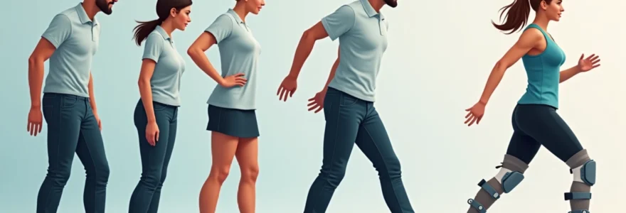
Physiotherapy and rehabilitation technology breakthroughs
The landscape of foot drop rehabilitation has been revolutionised by technological advances that enhance traditional physiotherapy approaches. Robotic-assisted devices now enable patients to practice proper gait patterns even when they lack the strength for independent movement, accelerating the rehabilitation process.
Functional electrical stimulation technology has become increasingly sophisticated, with devices now capable of detecting gait phases and delivering precisely timed stimulation. These systems can differentiate between swing and stance phases of walking, providing stimulation only when needed for foot clearance.
Virtual reality applications in rehabilitation allow patients to practice complex movements in safe, controlled environments. These systems can simulate challenging terrains and scenarios, building confidence whilst improving motor control. Studies show that VR-enhanced rehabilitation can improve outcomes by up to 25% compared to traditional methods alone.
The integration of technology with traditional rehabilitation approaches has transformed what’s possible for patients with foot drop. We’re seeing recoveries that would have been unimaginable even five years ago.
Biofeedback systems provide real-time information about muscle activation patterns, enabling patients to optimise their exercise technique. Surface EMG sensors can detect even minimal muscle activity, providing encouraging feedback during the early stages of recovery when visible movement may not yet be apparent.
Wearable sensors now enable continuous monitoring of gait parameters outside the clinic setting. These devices provide valuable data about walking patterns, step count, and progress over time, allowing rehabilitation teams to adjust treatment protocols based on real-world performance rather than clinic-based assessments alone.
Long-term functional mobility recovery assessments
Long-term follow-up studies reveal that meaningful recovery from foot drop can continue for years after initial injury, challenging traditional beliefs about neural recovery timelines. Research tracking patients over five-year periods shows continued functional improvements, particularly when combined with ongoing exercise programmes.
Functional outcome measures have evolved beyond simple strength testing to encompass quality of life, participation in activities, and psychological wellbeing. The comprehensive assessment approach recognises that successful rehabilitation extends beyond muscle function to include confidence, endurance, and social participation.
Recent studies indicate that patients who maintain active exercise programmes show significantly better long-term outcomes compared to those who discontinue structured activity after formal rehabilitation ends. This finding emphasises the importance of lifetime maintenance programmes for optimal functional preservation.
Community-based support programmes have emerged as crucial components of long-term success. Peer support groups, adaptive sports programmes, and ongoing education initiatives help patients maintain motivation and continue progressing years after their initial injury.
Technology integration continues to play an expanding role in long-term management, with smartphone applications now available for home exercise guidance and progress tracking. These tools enable patients to maintain structured rehabilitation programmes independently whilst staying connected with their healthcare teams for ongoing support and guidance.
 Good health cannot be bought, but rather is an asset that you must create and then maintain on a daily basis.
